Author: Nils Kohn
Affiliation(s): Donders Center of Medical Neurosciences, Radboud university medical centre
Research question(s):
- Are we able to replicate the four connectivity gradients of hippocampus-amygdala complex in the large Healthy Brain Data sample?
- What’s the relation between these connectivity gradients as established with HBS data and neurotransmitter maps within the complex?
- Lastly, we aim to improve our understanding of the connectivity gradients by asking, what are the behavioral and psychiatric meanings of these connectivity gradients?
Link: OSF Preregistration
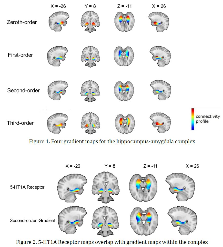
Abstract:The hippocampus and amygdala, as two adjacent medial temporal lobe structures, interact closely in human emotion and cognitive functions (Phelps, 2004; Richardson et al., 2004; Packard et al., 2021); their dysfunction also plays an important part in the development and maintenance of mental disorders like major depression, anxiety disorders (Boldrini et al., 2009; Shin & Liberzon, 2010). Revealing the precise functional role of the hippocampus and amygdala contributes to the targeted clinical application. So how hippocampal function changes along its long axis, and how amygdala interacts with this long-axis transition become the topic under much interest and debates (Strange et al., 2014). Animal anatomical studies showed dorsoventral (anterior-posterior) topographical gradients in hippocampal–cortical and subcortical connectivity (Witter, 1993; Kishi et al, 2006), while this organization in human has yet to be further examined (Strange et al., 2014).
Recently, an emerging connectivity analysis technique, the ‘connectopic mapping’ have become a promising tool for investigating this question in living human. Based on resting-state fMRI data, this method was designed to find several overlapping connectivity gradients within one brain region. Each connectivity gradient maps a varying topographic mode of connectivity changes within a pre-defined region-of-interest (ROI) in relation to other regions of the whole brain (Haak, Marquand, & Beckmann, 2018). By combining the hippocampus and amygdala into one complex, and utilizing the complex as the ROI, we will get the overview of hippocampal connectivity along the axis, and whether the amygdala maps into specific hippocampal regions. Furthermore, comparison with neurotransmitter maps and linking with behavioral and psychiatric domains will help us understand these data-driven derived gradients.
We have explored four connectivity gradients for the amygdala hippocampus complex (Figure 1) in a dataset of a psychiatric cohort (van Eijndhoven et al., 2022). The dominant gradient (zeroth-order) essentially shows changes following the coordinate space: starting from the amygdala then changing along the hippocampal long-axis. The second-order gradient maps are observed overlap with 5-HT1A Receptor maps bilaterally (left: fisher z = 1.955, p < 0.01; right: z = 1.843, p < 0.01; Bonferroni corrected. See Figure 2).
The above results are preliminary and unpublished. Based on our results from this highly comorbid sample, it is necessary and meaningful to examine whether these can be replicated in a dataset with healthy individuals. The comparison between healthy participants and patients will help us understand the generalizability and utility of this connectopic method, and the underlying mechanisms of mental disorders as well.